


INTRODUCTION
After traumatic injuries to teeth, such as luxation or avulsion, stabilization is of prime importance. Conditions for splints include:
- Passive, not active, force on a tooth
- Allowing for physiologic tooth movement
- No harm to soft tissue
- Not adversely affecting the occlusion
- Allowing for endodontic treatment to be performed if needed
- An easy-to-clean tooth
- Ease in removing from the tooth1,2
The most common treatment is the use of a flexible splint. Various materials are used, and the treatment is basically the same.3 Another treatment to stabilize the tooth after avulsion is suturing the tooth in place.4 Bleeding is often present, and the blood, as well as saliva, will make the adhesion between teeth and any resin difficult. Pediatric patients are often crying and fearful and can be challenging to manage through the sustained trauma of a long dental appointment. One must treat the traumatic tooth as quickly and concisely as possible.
Methods
This is the current protocol that we are using in these situations:
- Stabilize the tooth with light-cured resin (Light Fix [Sun Medical Co Ltd])
- Take an impression
- Create a vacuum-formed retainer (hard-type, 1.0-mm thickness)
- Deliver the retainer within 8 hours after trauma
- Keep the retainer on all day except when brushing teeth (28 days)
- Check the mobility of the tooth after 1, 7, 14, and 21 days
- In the case of luxation, taking an electric pulp test (EPT) (scale 1 to 10) after 1, 7, 14, and 21 days
- Checking the mobility of the tooth, percussion, palpation, and probing and taking an EPT and a periapical radiograph after 28 days
CASE REPORTS
Case 1
The patient was a 5-year-old girl who fell on top of a water bottle while drinking and avulsed the deciduous left lateral incisor. Her childcare worker washed the tooth with tap water and cleaned the dirt off the tooth, removing the periodontal membrane. The tooth was replanted and stabilized by a light-cured resin, and an impression was taken. She returned to the dental clinic 3 hours later and received a vacuum-formed retainer. Her mother was instructed that her daughter should wear the retainer all day except when brushing her teeth. The tooth was checked after 1, 7, 14, and 21 days, and a dental radiograph was taken after 28 days. Endodontic treatment was performed after the tooth was stable. A 12-month followup was within normal limits (Figure 1).
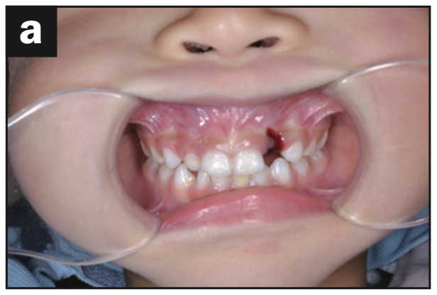
Figure 1a. Preoperative intraoral photograph.
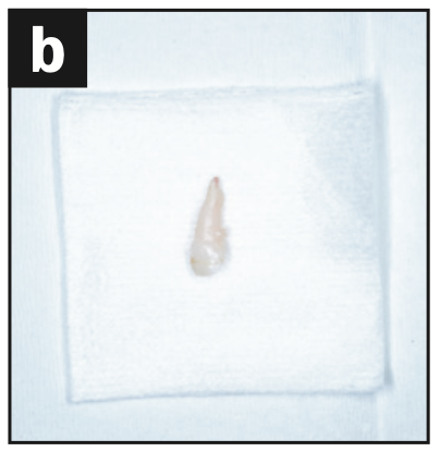
Figure 1b. Avulsed tooth
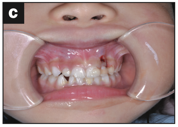
Figure 1c. Intraoral photograph; the tooth was replanted and fixed by light-cured resin.
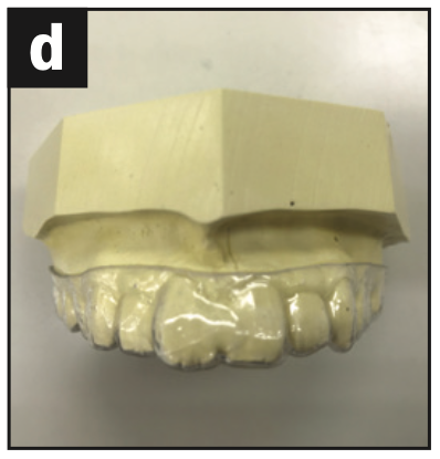
Figure 1d. Working model with a vacuum- formed retainer.
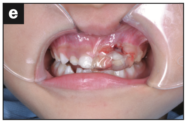
Figure 1e. Intraoral photograph wearing the vacuum-formed retainer.
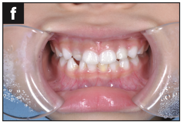
Figure 1f. Intraoral photograph at 28 days postoperative.
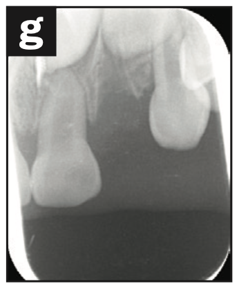
Figure 1g. Pre-op radiograph.
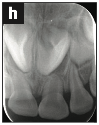
Figure 1h. Radiograph immediately after replanting.
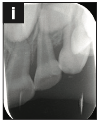
Figure 1i. Radiograph at 28 days post-op.
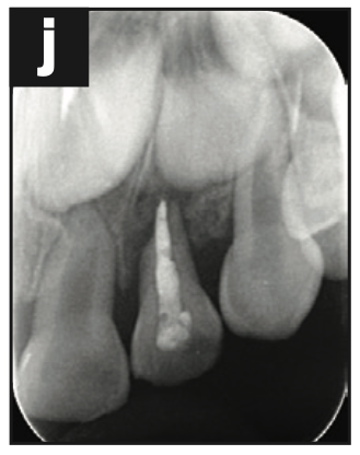
Figure 1j. Radiograph of root canal filling.
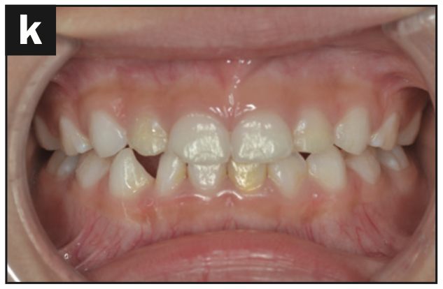
Figure 1k. Intraoral photograph at 12 months post-op.
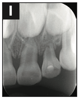
Figure 1l. Radiograph at 12 months post-op.
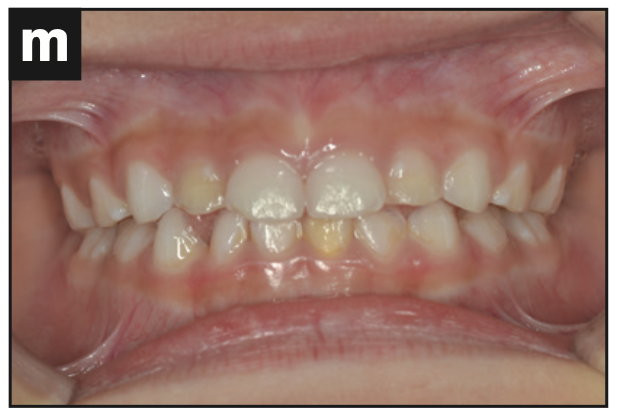
Figure 1m. Intraoral photograph at 18 months post-op.
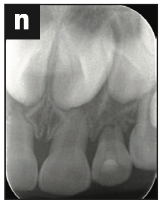
Figure 1n. Radiograph at 18 months post-op.
Case 2
This patient was a 24-year-old female. She had fallen, causing lateral luxation of her maxillary left central incisor. The next morning, she came to the office, where it was stabilized by a light-cured resin, and an impression was taken. Six hours later, she returned and received a vacuum-formed retainer. She was instructed to wear the retainer all day except when brushing her teeth. Sensitivity tests were performed after 1, 7, 14, and 21 days. Mobility of the tooth and palpation were checked, an EPT was done, and a dental radiograph was taken after 28 days. The tooth had +1 mobility in the labial-palatal direction and no reaction to the EPT. Three months later, the tooth was restored and responded to an EPT (8/10). At the 4-month recall, the tooth responded to EPT (4/10). At 5 months, a sinus tract was noted. The tooth did not respond to the EPT. A lesion at the apex was seen on the radiograph. The diagnosis was pulp necrosis, and root canal treatment was performed in 2 visits. One month later (6 months after the trauma occurred), the sinus tract disappeared, and the radiograph showed the process of healing (Figure 2).
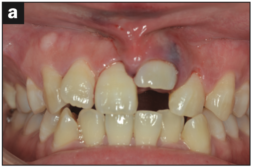
Figure 2a. Pre-op intraoral photograph of displacement.
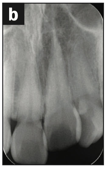
Figure 2b. Pre-op radiograph of displacement.
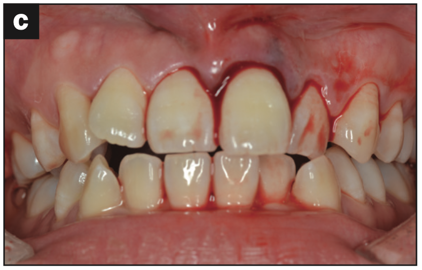
Figure 2c. Intraoral photograph of the replanted tooth.
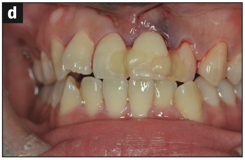
Figure 2d. Intraoral photograph. The tooth was fixed with light-cured resin.
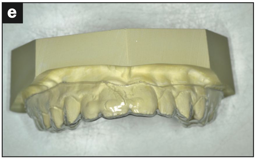
Figure 2e. Working model with a vacuum-formed retainer.
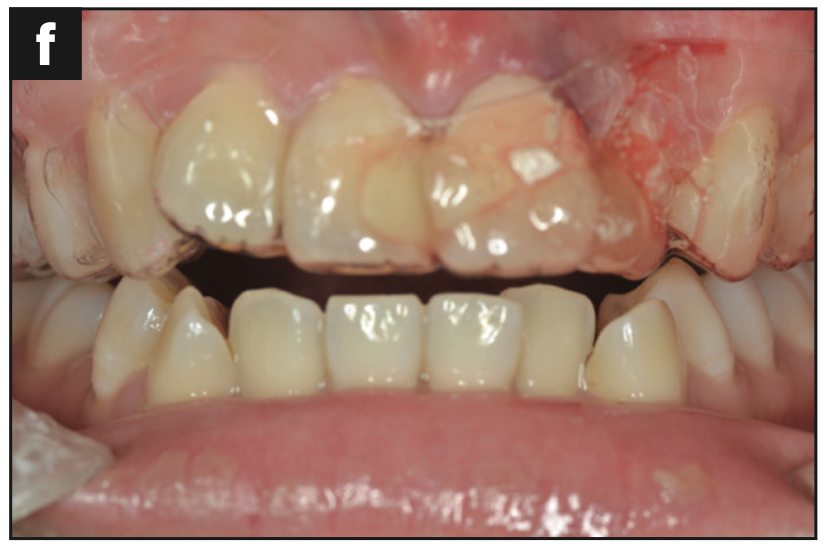
Figure 2f. Intraoral photograph. Wearing a vacuum-formed retainer.
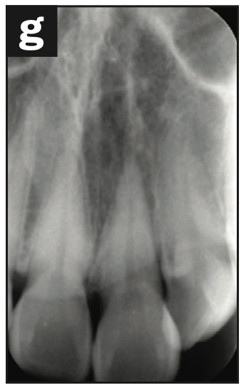
Figure 2g. Radiograph immediately after replanting.
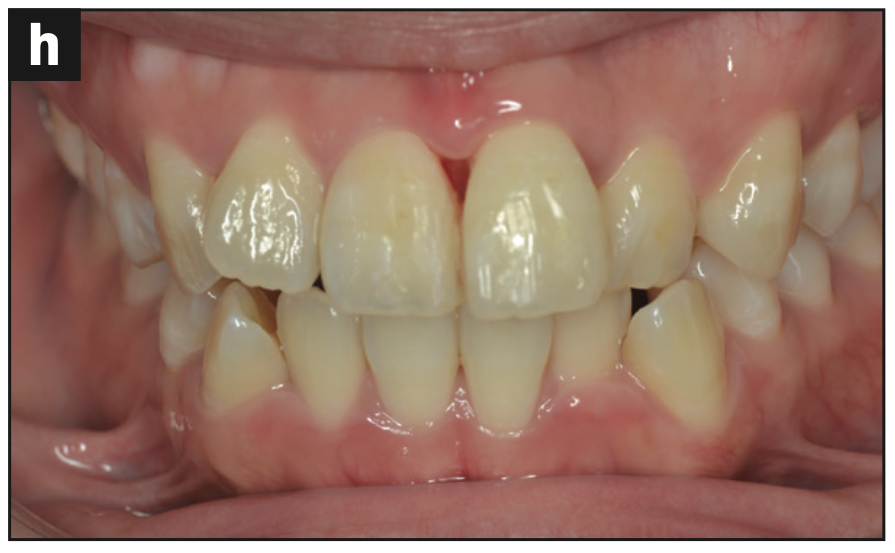
Figure 2h. Intraoral photograph at 28 days post-op.
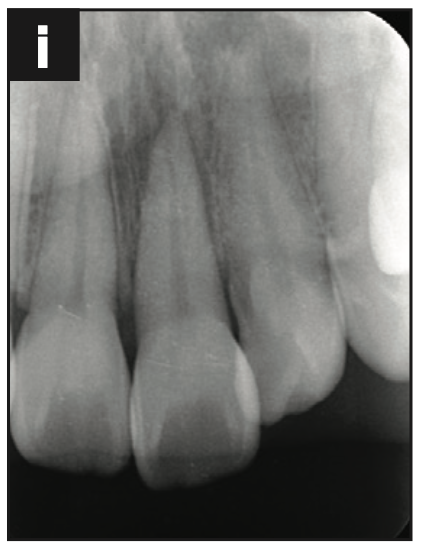
Figure 2i. Radiograph at 28 days post-op.
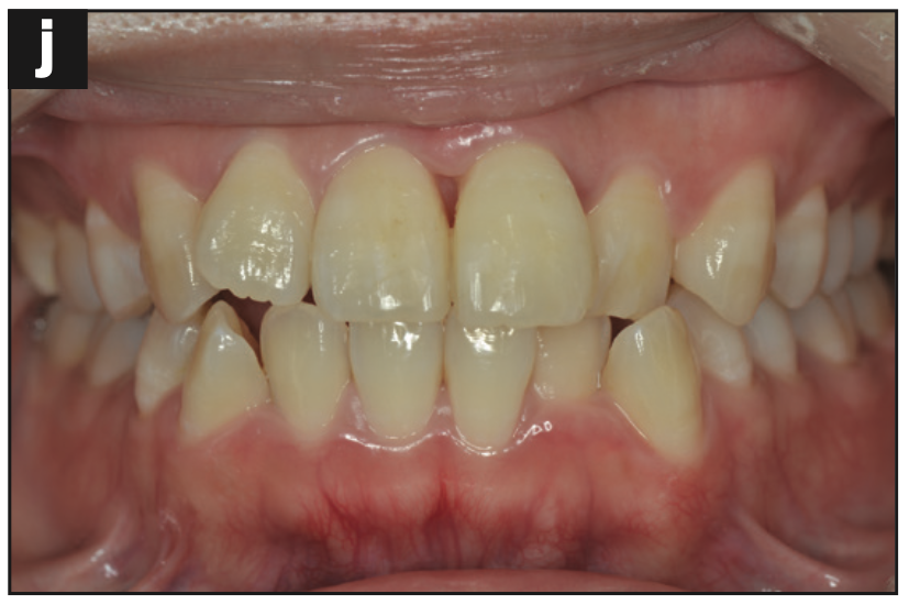
Figure 2j. Intraoral photograph at 3 months post-op.
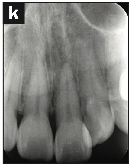
Figure 2k. Radiograph at 3 months post-op.
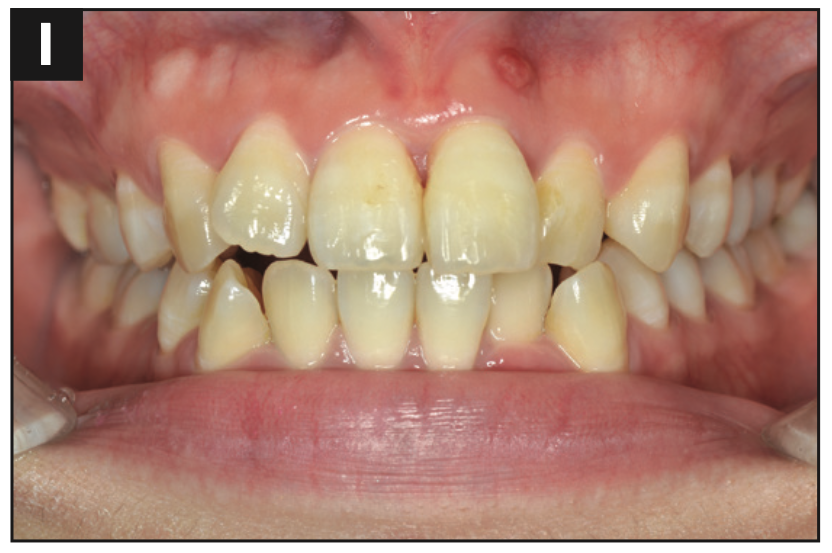
Figure 2l. Intraoral photograph at 5 months post-op; the sinus tract was present.
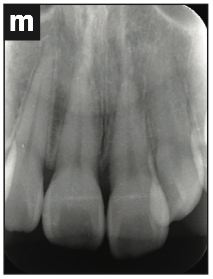
Figure 2m. Radiograph at 5 months post-op; the apex of the tooth had a lesion.
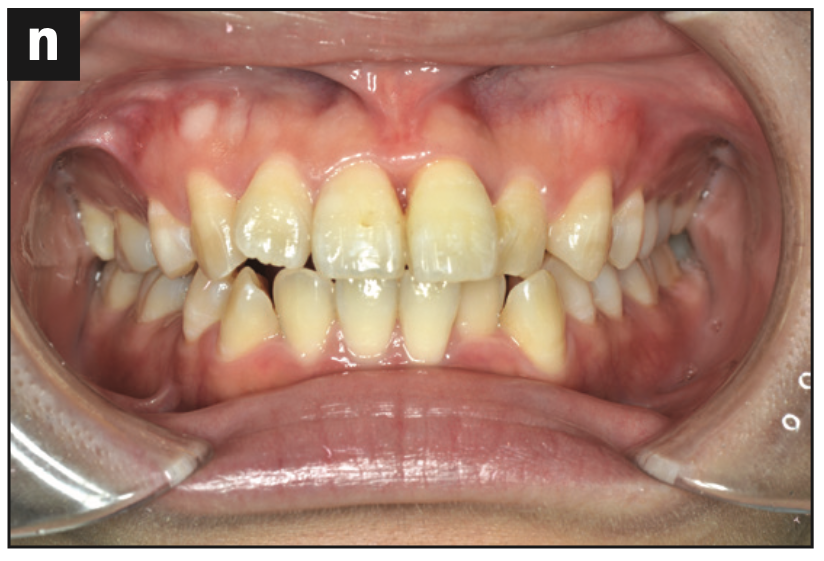
Figure 2n. Intraoral photograph at 6 months post-op; the sinus tract was gone.
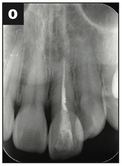
Figure 2o. Radiograph at 6 months post-op; the apex of the tooth shows healing.
Case 3
A 42-year-old female fell, causing palatal luxation of her maxillary left central incisor. An alveolar bone fracture on the palatal side was seen in the CBCT. The fracture was reduced and stabilized by a light-cured resin, and an impression was taken. Two hours later, she returned and was given a vacuum-formed retainer. She was instructed to wear the retainer all day except when brushing her teeth. It was notable that she had a tongue-thrusting habit. Sensitivity tests were performed after 7 and 28 days. Mobility of the tooth, an EPT, palpation, and a dental radiograph were taken after 28 days. At the 28-day followup, there was slight mobility of the tooth and no reaction to the EPT. At the 2-month followup, the tooth was not discolored, had no mobility, was negative to EPT, and was slightly sensitive to percussion. There was no periapical lesion on the radiograph and no sinus tract. At the 3-month followup, the tooth was responsive to EPT (9/10) and had slight percussion pain. At the 4-month followup, the tooth was responsive to EPT (9/10) and no longer percussion-sensitive (Figure 3).
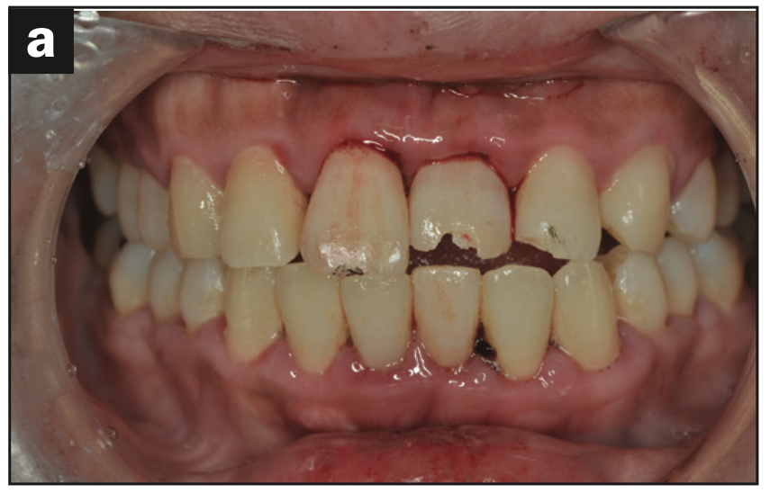
Figure 3a. Pre-op intraoral photograph of displacement.
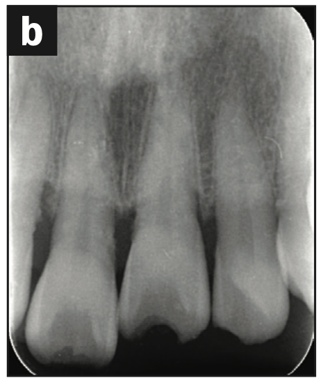
Figure 3b. Pre-op radiograph of displacement.
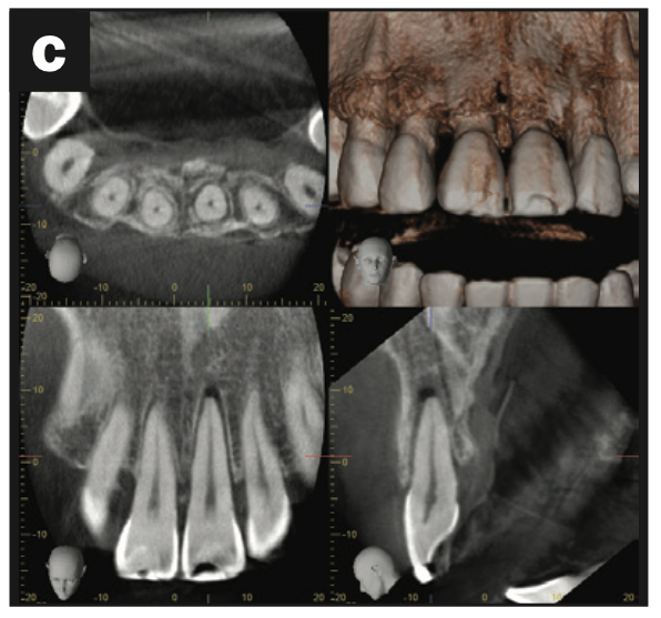
Figure 3c. CBCT scan of the alveolar fracture of the palatal bone.
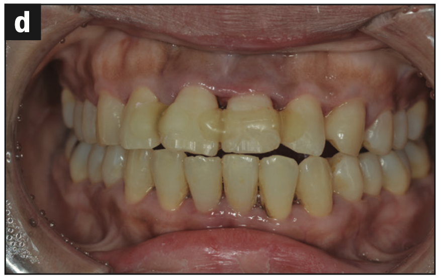
Figure 3d. Intraoral photograph. The tooth was replanted and fixed by light-cured resin.
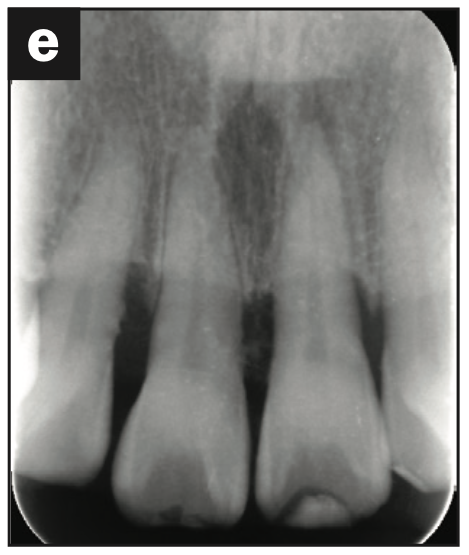
Figure 3e. Radiograph immediately after replanting.
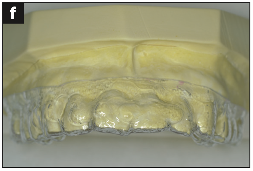
Figure 3f. Working model with a vacuum-formed retainer.
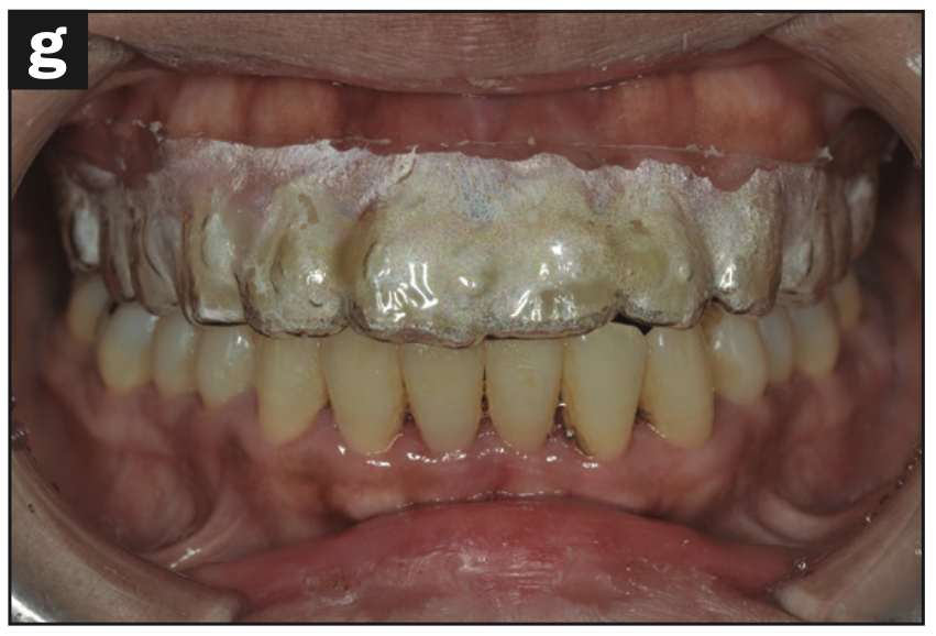
Figure 3g. Intraoral photograph. Wearing a vacuum-formed retainer.
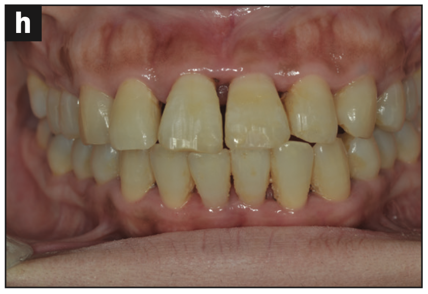
Figure 3h. Intraoral photograph at 28 days post-op.
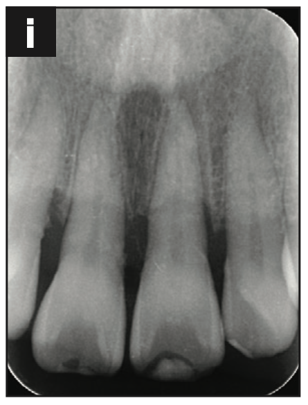
Figure 3i. Radiograph at 28 days post-op.
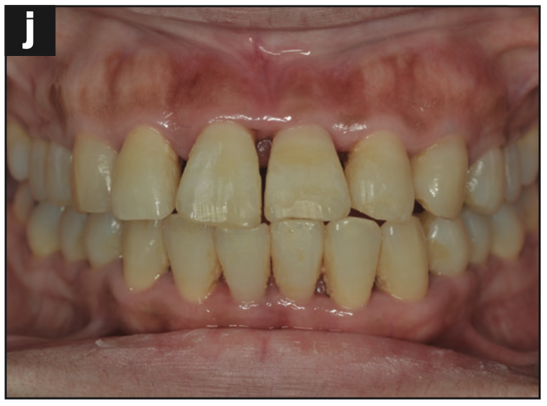
Figure 3j. Intraoral photograph at 3 months post-op.
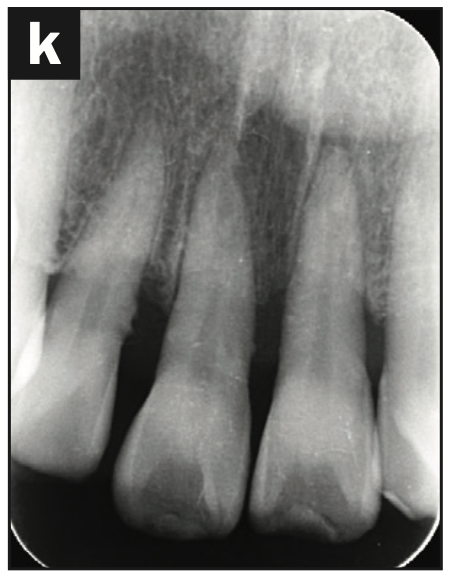
Figure 3k. Radiograph at 3 months post-op.
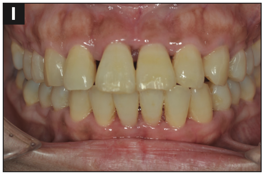
Figure 3l. Intraoral photograph 4 months post-op.
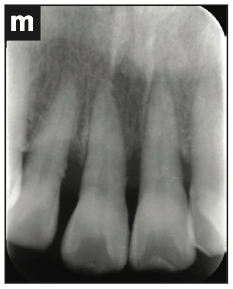
Figure 3m. Radiograph at 4 months post-op.
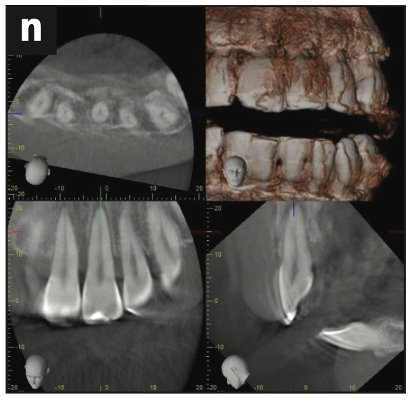
Figure 3n. CBCT scan at 4 months post-op.
DISCUSSION
Trauma patients most often will come to the dental office in an emergency. They are often young patients who are frightened and have difficulty understanding what we are doing as well as following our instructions. Our goals as treating dentists are to remove any contaminants from the oral cavity and restore and stabilize the luxated, subluxated, or avulsed teeth. It may be difficult at the emergency appointment to establish a proper bond with resin, but it is critical to take an impression in order to create a vacuum-pressed retainer. Ideally, a digital impression could be performed, which will significantly decrease the chances of further trauma to the teeth. The patient and guardian/parent are instructed to always wear the vacuum-formed retainer except when brushing the teeth for a period of 28 days. The pathological healing process of subluxation, luxation, and avulsion stabilized by the vacuum-formed retainer is the same as using a flexible splint.5,6
The patient in case 1 was young. She ignored instructions and touched her teeth, removing the resin. The patients of cases 2 and 3 were adults and followed the instructions very well. Adult patients may not require 28 days of fixation.
CONCLUSION
Traumatic injuries such as luxation and avulsion stabilized by vacuum-formed retainers (thickness of 1.0 mm) show healing after 28 days if worn constantly except when brushing the teeth.
ACKNOWLEDGMENTS
Dr. Tsumori would like to thank the staff members of Meiwa Dental Clinic in Japan, Dr. Samuel Kratchman, and Dr. Yoshi Terauchi.
REFERENCES
1. Andreasen JO, Andreasen FM. Essentials of Traumatic Injuries to the Teeth: A Step-by-Step Treatment Guide. 2nd edition. Mosby; 2000.
2. American Academy on Pediatric Dentistry Council on Clinical Affairs. Guideline on management of acute dental trauma. Pediatr Dent. 2010;32(6 Supple):202–12.
3. Berman LH, Hargreaves KM. Cohen’s Pathways of the Pulp. 12th edition; Elsevier; 2021.
4. Gupta S, Sharma A, Dang N. Suture splint: an alternative for luxation injuries of teeth in pediatric patients—a case report. J Clin Pediatr Dent. 1997;22(1):19-21.
5. Sobczak-Zagalska H, Emerich K. Best Splinting Methods in Case of Dental Injury—A Literature Review. J Clin Pediatr Dent. 2020;44(2):71–8. doi:10.17796/1053-4625-44.2.1
6. Baruch H, Ehrlich J, Yaffe A. [Splinting—a review of the literature]. Refuat Hapeh Vehashinayim (1993). 2001;18(1):29-40, 76.
ABOUT THE AUTHORS
Dr. Tsumori graduated from Tokyo Dental College in 2003, then served as a clinical assistant at Tokyo Dental College from 2003 to 2004. He is the owner of Meiwa Dental Clinic. He can be reached by contacting Dr. Samuel Kratchman at sikratch@comcast.net.
Dr. Kratchman received a BS in Biology and his DMD degree at Tufts University in Boston and his certificate in endodontics from the University of Pennsylvania, where he is an associate professor of endodontics, the assistant director of graduate endodontics, and in charge of microsurgery. Dr. Kratchman co-authored the textbook Microsurgery in Endodontics with Dr. Syngcuk Kim in 2017 and developed a patented instrument called the S-Kondenser for obturation of root canals. Dr. Kratchman lectures nationally and internationally, in addition to maintaining 4 private practices in Exton, West Chester, Paoli, and Bryn Mawr, Pa. He can be reached at sikratch@comcast.net.
Disclosure: The authors report no disclosures.


