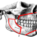
The sagittal split osteotomy is a widely used procedure for the correction of dentofacial deformities. Traditionally it has been suggested that lower third molars (3Ms) should be extracted at least 6 months prior to corrective surgery because of an increased risk of complications. However, recent studies have removed 3Ms concomitant with the sagittal split osteotomy procedure and there is a debate regarding the appropriate timing for removal of 3Ms and the impact on complications during sagittal split osteotomy.
The aim of this review was to investigate whether the presence of third molars during sagittal split osteotomy of the mandible increases the risk of complications.
Methods
A protocol for the review was registered on the PROSPERO database. Searches were conducted in the Cochrane Central, Medline/PubMed, LILACS, Scopus, Dentistry and Oral Sciences Source (DOSS) and OpenGrey database with no limits on publication dates or language. Randomised clinical trials (RCTs) and observational studies (prospective and/or retrospective) evaluating the occurrence of complications during sagittal split osteotomy were considered. Two reviewers independently selected studies with data being extracted by one reviewer and crosschecked by a second. Risk of bias was assessed using the Newcastle–Ottawa Scale (NOS). Meta-analyses were conducted, and overall certainty of evidence assessed using the Grading of Recommendations, Assessment, Development, and Evaluation (GRADE) approach.
Results
- 15 studies (11 retrospective, 4 prospective) involving a total of 3909 patients were included.
- Studies were from 9 countries, USA (5 studies) Canada (3 studies, Netherlands (2 studies) one each from Iran, South Africa and Taiwan and one multi-centred study from the Netherlands, UK, Germany, and Spain.
- 13 studies were considered to be at high risk of bias and 2 a low risk.
- Of 7651 mandibular sagittal split osteotomies, 3272 (42.8%) involved a 3Ms at the surgical site, while 4379 (57.2%) did not.
- 11 studies reported on the surgeon’s profile and 4 studies reported on surgery duration.
- 670 complications were reported in the 14 studies included in the meta-analyses. Inferior alveolar nerve (IAN) exposure in the proximal segment was the most frequent complication (n = 409), followed by bad splits (n = 151).
- Meta-analyses showed no significant differences in complications in presence of 3Ms (see table)
| Complication | No. of studies | Risk ratio (95%CI) |
| Nerve exposure | 4 | 1.24 (0.70 to 2.19) |
| Neurosensory disorders | 3 | 0.46 (0.21 to 1.00) |
| Postoperative infection | 1 | 1.54 (0.86 to 2.78) |
| Osteosynthesis remove | 1 | 0.61 (0.20 to 1.83) |
- Sub-group analyses suggested an increase in bad splits associated with 3Ms in studies up to 150 3Ms but not with >150 (see table).
| No. of studies | Risk ratio (95%CI) | |
| < 150 cases | 6 | 2.54 (1.15 to 5.63) |
| >150 cases | 7 | 0.93 (0.42 to 2.08) |
| Overall | 13 | 1.45 (0.79 to 2.69) |
- Subgroup analyses showed an increased risk of bad splits in the presence of 3Ms in European and USA studies (see table).
| Region | No. of studies | Risk ratio (95%CI) |
| European | 4 | 1.84 (1.10 to 3.08) |
| USA | 4 | 4.71 (1.92 to 11.52) |
| Canadian | 3 | 0.53 (0.29 to 0.98) |
| Africa/Asia | 2 | 1.47 (0.03 to 62.30) |
| Overall | 13 | 1.45 (0.79 to 2.69) |
- Location of bad splits was not associated with the presence of 3M (see table)
| Bad Splits | No. of studies | Risk ratio (95%CI) |
| Buccal cortex | 1 | 0.08 (0.01 to 1.29) |
| Lingual plate | 2 | 2.86 (1.36 to 6.00) |
| Proximal | 9 | 0.83 (0.64 to 1.08) |
| Distal | 8 | 1.29 (0.92 to 1.82) |
- The GRADE assessment of the certainty of the evidence was very low.
Conclusions
The authors concluded: –
There was no statistically significant relationship between the presence of 3Ms and complications such as the presence of a nerve in the proximal segment of the osteotomy, infections, surgical removal of osteosynthesis material, and the occurrence of bad splits. However, the quality of evidence for this is very low. Thus, we strongly recommend future investigations with detailed methodologies and well- described surgical protocols to provide higher quality evidence to surgeons concerning this topic.
Comments
The reviewers have searched a good range of databases for studies addressing their question. 15 studies were identified involving a reasonable number of patients however there were all an observational design with a majority (11 studies) being retrospective in nature. In addition, 13 of the 15 studies were assessed as being at a high risk of bias with the review authors also highlighting methodological variation involving patient data selection and measurement and incomplete data. So, while the findings suggest no significant relationship between the presence of 3Ms and osteotomy complications the limited quality of the included studies means there is very low certainty in the findings. Further high quality well-reported prospective studies are needed to address this topic.
Links
Primary Paper
Cetira Filho EL, Sales PHH, Rebelo HL, Silva PGB, Maffìa F, Vellone V, Cascone P, Leão JC, Costa FWG. Do lower third molars increase the risk of complications during mandibular sagittal split osteotomy? Systematic review and meta-analysis. Int J Oral Maxillofac Surg. 2022 Jul;51(7):906-921. doi: 10.1016/j.ijom.2021.12.004. Epub 2021 Dec 23. PMID: 34953646.
Other references
Dental Elf – 7th Mar 2012
Presence of mandibular third molars during sagittal split osteotomies did not increase complications
Picture Credits
By Henry Grey – Henry Gray (1918) Anatomy of the Human Body (See “Book” section below), Public Domain, Link
