
Zirconia puzzle: what makes zirconia unique and how to choose the right zirconia
Zirconia may be part of your everyday vocabulary, but how much do you know about this ceramic material? Learn more…


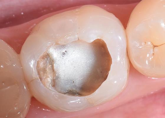
How often do you place liners under your restorations? While you may not use them as often as you used to, liners can still be very useful in the modern dental practice. Discover how liners and liner materials have evolved, their benefits, and how you can make the most of them.
Historically, cavity preparations were much more aggressive, leading to the exposure of deep dentin. Liners were indicated to protect the pulp from thermal and galvanic stimuli conducted through metal-based restorations and chemical irritants (restorative materials) and to induce tertiary dentin formation.
However, modern composites have largely supplanted the use of amalgam, and cavity preparations have evolved according to minimally invasive concepts, which has changed how we use liners. Even with the advent of advanced adhesive solutions, several materials are still in use as liners, but for different reasons. In this article, we will discuss the clinical circumstances where using a liner may be beneficial, how the use of liners has changed over time, the restorative materials dentists have to choose from, and what each option brings to the table.
Traditionally, clinicians were taught to remove all carious tissue prior to placing restorations, which, in deep carious lesions, could lead to very thin layers of dentin between the cavity floor and the pulp. In a worst-case scenario, removing carious tissue can lead to unintended pulp exposure, which may require pulp capping. However, this aggressive treatment style has fallen out of favor as more conservative strategies have emerged that preserve tooth structure, the ultimate goal being to protect the pulp.
In recent years, it has become ever more accepted – thanks to robust scientific evidence – that the best way to treat carious dentin is by using selective carious tissue removal. In deep cavities of vital teeth, with a positive response to sensitivity tests, leathery or even soft dentin can be left on the cavity floor so as not to damage the pulp or risk pulpal exposure. Retaining dentin as a barrier between the restorative material and the pulp has reduced the need for liners in deep cavities for a therapeutic or “protective” reason. Maintaining a thicker dentin layer between the cavity floor and pulp and modern adhesives’ ability to seal caries-affected dentin enables this technique (fig 1).
In very deep restorations, dentists may not always feel comfortable placing an adhesive directly. Instead, a more biocompatible material such as a resin-modified glass ionomer liner (RMGI) is recommended to help protect against post-operative sensitivity. It is important to note that despite the available evidence on selective caries removal, some dental schools are still teaching complete (nonselective) caries removal strategies. Caries removal strategies also vary among clinicians. Even when following selective caries removal techniques, the pulp may still be unintentionally exposed in very deep regions of the dentin. In that case, a direct pulp capping agent may be indicated.

Figure 1: Deep caries lesion restored following the selective removal approach. Clinical pictures were performed in collaboration with Prof. Dr. Rayssa Ferreira Zanatta.
A. An asymptomatic, vital tooth that reacted normally to a sensitivity test to cold stimuli, presenting a deep lesion (radiolucency at the inner third/inner fourth of dentin)
B. Margins and peripheral walls of the cavity prepared until sound tissue/hard dentin.
C. Deepest carious dentin towards the pulp at the cavity floor is preserved to avoid pulp exposure or an excessively thin dentin layer between cavity and pulp.
D. In a closer view it is possible to observe the layer of soft carious dentin that was not removed.
E. The rationale of selective caries removal is to preserve a layer of dentin at the cavity floor and avoid unnecessary trauma or complications to the pulp that usually occur when all carious dentin is removed. With well-sealed restorations (good adhesive procedures on margins and peripheral walls), any microorganisms left become unviable, and the lesion does not progress.”1
There have been great advancements in dental adhesive technology in recent decades, and there are now a number of effective and clinically proven strategies to promote adhesion to tooth substrates.
Many of today’s mild self-etch and universal adhesives also incorporate functional monomers like 10-MDP that promote a chemical bond to the calcium in dentin’s hydroxyapatite – which is preserved in the hybrid layer due to the adhesive’s mild acidity. This chemical interaction might cooperate for the longevity of the adhesion. However, since enamel, which has a much higher mineral content than dentin, still benefits from a prior phosphoric acid etching, the selective enamel etch technique is the most recommended approach when using self-etch or universal adhesives. When one uses the selective caries removal technique correctly, and a thicker layer of dentin is preserved between the cavity floor and the pulp, you can use an adhesive directly on this substrate. In these cases, materials like 3M™ Scotchbond™ Universal Plus Adhesive, a universal adhesive, have mild acidity and functional monomer 10-MDP to create a good seal without needing any lining materials (fig 2). However, with all of this in mind, adhesives should never be in direct contact with the pulp or areas too close to pulpal tissue (deep cavities where non-selective caries removal was performed or too much dentin has been unintentionally removed). Which raises the question – what can you use in these circumstances?

Figure 2: Restorative procedures of the clinical case presented on Figure 1. Clinical pictures performed in collaboration with Prof. Dr. Rayssa Ferreira Zanatta.
A. Selective enamel etch with phosphoric acid.
B. Application of a universal adhesive system (3M™ Scotchbond™ Universal Plus).
C. A thin layer of flowable composite resin (3M™ Filtek™ Supreme Flowable Restorative) is applied and adapted to the cavity walls using a probe. The main reason for using a flowable is to easily achieve good composite adaptation to the adhesive layer, particularly at uneven areas and sharp angles of the cavity.
D. Restoration is performed using a regular viscosity composite. In this case, a bulk-fill composite (3M™ Filtek™ One Bulk Fill Restorative) was used and finalized with brown stains at the occlusal sulcus.1
When you need a liner, selecting the right material is key. The ideal material is biocompatible, adapts well to the cavity walls and floor, delivers good adhesion, has very low solubility, and is easy to apply. Liners should be applied in a localized, thin layer, so the material must be easy to place – particularly when it comes to deep cavities.
Glass ionomers or resin-modified glass-ionomers, such as 3M™ Vitrebond™ Plus Light Cure Glass Ionomer Liner/Base, are a popular choice as they can be used to line deep cavity preparations or where needed to reduce post-operative sensitivity. Plus, these materials can be used to create a smooth cavity floor over direct pulp capping materials (like calcium hydroxide, MTA, and others) especially when the latter is soluble or does not adhere to dentin.
In posterior restorations, many clinicians use a flowable composite as the first restorative layer after the adhesive. While not required, flowable may help improve adaptation of the final composite to the cavity walls (especially when there are sharp angles in the cavity preparation) and restorative matrices (fig 3). Some clinicians and researchers advocate that a first layer of flowable composite may act as a “stress-absorber” under more rigid restorative materials due to its lower elastic modulus. This increasing indication for flowable composites also encompasses restorative steps known as “immediate dentin sealing,” “resin coating” (primarily used for indirect procedures), and the “snowplow” and “injection molding” techniques. It is important to note that the flowable composite itself is not actually a liner – it doesn’t have the traditional connotation of biocompatibility or pulp protection – but as a step in achieving a good dentin seal and marginal adaptation.

Figure 3: Schematic illustration of the case presented in Figures 1 and 2.
A. Illustration of deep carious lesion in an asymptomatic vital premolar.
B. Radiograph of the deep carious lesion with asymptomatic vital premolar.
C. Selective caries removal (margins and peripheral walls on sound tissue and/or hard dentin), preserving some of the deepest carious dentin.
D. After selective etching of the enamel margins, a universal adhesive is applied and light-cured, followed by a thin layer of a flowable composite to provide good adaptation to the cavity walls. The application of the flowable composite is not mandatory, but a preferred technique by many practitioners due to its reduced viscosity and ease of application.
E. The cavity is restored using the professional’s preferred protocol, either with a multi-layered or a bulk-fill composite.1
When selecting the best material for each case, it’s important to review your options and keep clinical history in mind. For posterior restorations, I like using an adhesive such as 3M™ Scotchbond™ Universal Plus Adhesive, which has a dentin-like radiopacity. This radiopacity is especially helpful when utilizing the selective caries removal technique (fig 4). According to in vitro studies, it also offers better adhesion to caries-affected dentin, like to the cavity floor after selective caries removal.
For the first layer in many posterior restorations, I use a thin layer of a flowable material (either 3M™ Filtek™ Supreme Flowable Restorative or 3M™ Filtek™ Bulk Fill Flowable Restorative), for their excellent adaptation, ease-of-use, and versatile delivery systems. Both flowables have ergonomic syringes with bendable cannulas that improve access to the cavity – making them ideal for precise placement.

Figure 4: Selective carious tissue removal with a hand excavator (Photo courtesy of Dr. Michal Dekel-Steinkeller)1
In cases where we may have removed a little too much carious tissue and are near the pulp – where the bottom of the prep appears pinkish, almost like you can see the pulp through very thin dentin – it’s advisable to place a more biocompatible material as a liner. Vitrebond Plus is an example of an RMGIC that provides the biocompatibility we need, along with good adaptation and adhesion (fig 5 and 6).

Figure 5: In clinical cases with deep carious lesions (vital, positive response to a pulp sensitivity test) that are subject to a nonselective caries removal, or in those where an excessive removal occurs leading to a very thin dentin on the cavity floor, liners with better biocompatibility than adhesives are still required. Clinical picture performed in collaboration with Prof. Dr. Fernando Vilain de Melo.
A. Example of a deep caries lesion that was removed up to very close to the pulp.
B. See the pinkish axial wall, denoting a very thin dentin layer.
C. Schematic illustration of nonselective caries removal leaving a very thin dentin layer over the pulp.
D. A thin layer of resin-modified glass-ionomer cement with good biocompatibility (such as 3M™ Vitrebond™ Plus Light Cure Glass Ionomer Liner/Base) is indicated on very deep cavity floors when there is no pulp exposure.
E. The restorative technique after the liner follows the same principles presented in Figures 2 and 3.1

Figure 6: 3M™ Vitrebond™ Plus is placed directly on caries-affected dentin (Photo courtesy of Dr. Michal Dekel-Steinkeller)1
The adoption of conservative caries removal approaches and the evolution of adhesive techniques has reduced the need for liners under composite restorations. However, there are still occasions where a liner material may be helpful or even critical to a restoration. Glass ionomers and resin-modified glass ionomers, for example, have a long history as liners and are still indicated due to their biocompatibility and adhesive properties. Flowable composites can be used as a first restorative layer after the adhesive because of their ability to adapt to uneven cavity floors, walls, and sharp angles, as well as their versatile delivery systems.

Zirconia may be part of your everyday vocabulary, but how much do you know about this ceramic material? Learn more…
Achieving the correct shape is vital to a natural-looking anterior composite restoration. Learn how the 3M™ Filtek™ Matrix, paired with…
Direct composite restorative procedures can be challenging, particularly when it comes to esthetic cases. Discover how new techniques and tools…