
By: Jeffrey W. Horowitz, DMD, FAGD, D-ABDSM, D-ASBA
Have you ever run into pitting and erosive tooth lesions during your time as a dentist?
Here is a case study on my experience with it.
Pitting and Erosive Tooth Lesions: Case Study
Patient Information:
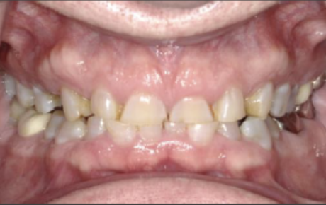
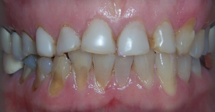
- 69-year-old white male presents for a dental exam concerned with tooth wear and minor orthodontic relapse
- Medical history positive for hypertension, G.E.R.D., and Hypothyroidism
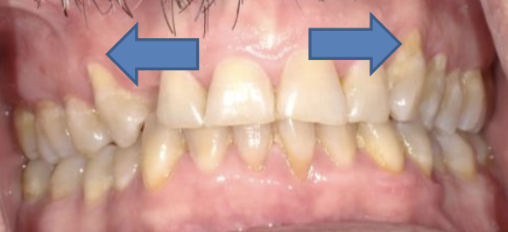
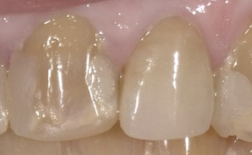
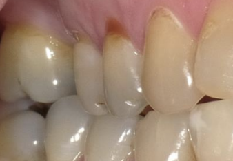
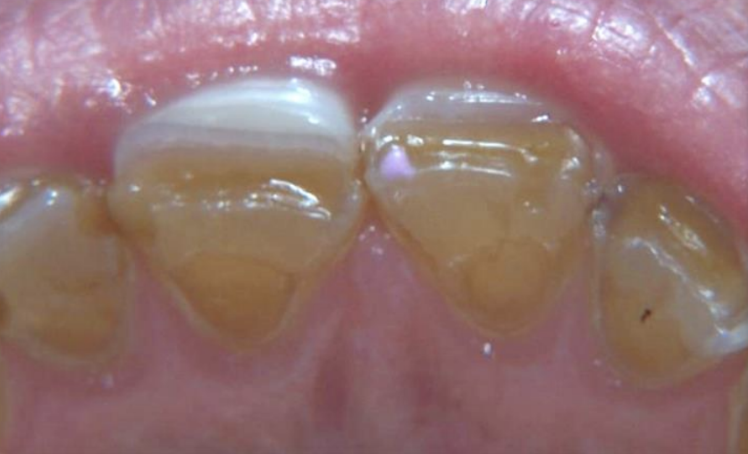
- Dental/Perio Exam negative other than Amalgam leakage #7, posterior erosive pitting lesions, and loss of lingual enamel 8,9
- Archform is normal but a deep bite exists
- Soft tissue exam positive for oro- pharyngeal inflammation/edema
Etiology of Tooth Structure Loss
Tooth erosion happens when acids wear away the enamel on teeth. Here are some common causes of tooth erosion and pitting:
1. Attrition
Tooth to Tooth
2. Abrasion
Extrinsic etiology; toothbrush, the particle size of dentifrice, abrasive diet, etc.
3. Abfraction
From physical forces, flexure, fracture
4. Erosion
Chemical structural breakdown
5. Infectious
Caries
Many Lesions are Multi-Factorial, but Which Came First?
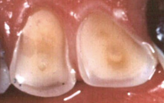
Non-Carious “pitted” or “cupped” lesions on the occlusal and lingual surfaces; Think acid insult from intrinsic source (reflux/bulimia. Undiagnosed Apnea is a common source of reflux and attrition.
If the structural loss on the occlusal surface does not correspond to the opposing tooth structure (the models don’t fit), rule out attrition.
Most facial lesions are multi-factorial. True abfractions are typically seen with functional pre-maturities but exposed dentin is now more susceptible to both erosion (extrinsic and intrinsic sources) abrasion and potential caries.
Differential Diagnosis
Attrition: The patient has anterior prematurities, lingual 8,9, but posterior lesions are too well-defined to suggest tooth-to-tooth wear. Models do not correspond.
Abrasion: Not likely, lesions too isolated to be caused by an extrinsic force.
Abfraction: None noted
Erosion: Most likely. No facial lesions were noted suggesting intrinsic origin versus dietary.
Infection (Caries): None noted except #7 amalgam
Pitting and Erosive Tooth Lesions: Summary
The patient was confirmed to have sleep apnea following a home sleep study diagnosed by a board-certified sleep physician.
Primary Diagnosis: Acid Erosion (Intrinsic origin) secondary to O.S.A. Primary erosion 8,9 with hyper-occlusion secondary to orthodontic treatment accelerating structural breakdown.
G.E.R.D. and oral pH controlled by treating OSA with Oral Appliance and OTC Prilosec.
Anterior occlusion (Hyper-occlusion on 8,9) & crowding were corrected with clear aligner therapy.
Recommendation for Ultra Soft toothbrush and low abrasive toothpaste (R.D.A. < 40).
No treatment of lesions as the patient had low caries risk. Will monitor.
Up Next: Scalloped Tongue, What Does it Mean?

